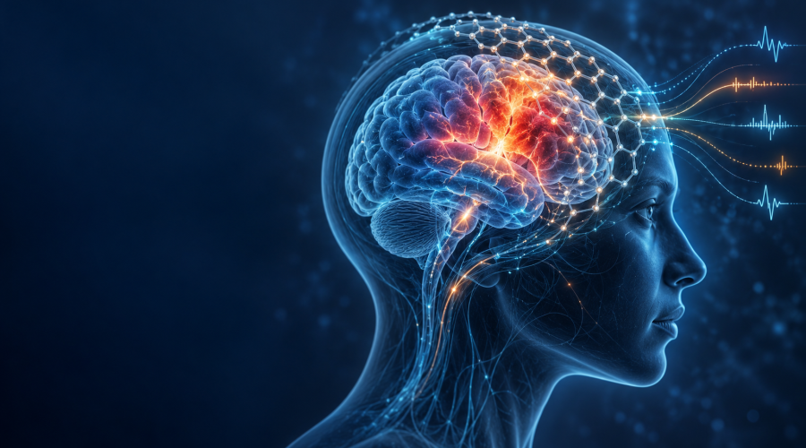
Graphene Sensors Could Help Map Stroke Damage in Real Time
Usagevpn.com – A new graphene-based monitoring system has enabled scientists to observe harmful waves of brain activity after stroke with exceptional detail, offering a possible route toward more targeted treatments in the future.
The technology was tested in mice by an international research group involving Spain’s National Research Council (CSIC), the University of Manchester, the Catalan Institute of Nanoscience and Nanotechnology (ICN2), and German company Multi Channel Systems. Their work appeared in the scientific journal Brain.
Researchers focused on ischaemic stroke, which occurs when a blockage cuts off blood supply to part of the brain. The immediate shortage of oxygen and nutrients can injure brain cells, but the initial blockage is not always the end of the damage. In the surrounding tissue, slow-moving disruptions in electrical activity can travel through the cortex and contribute to the expansion of lesions.
Tracking damaging electrical waves
These disturbances are known as spreading cortical depolarisations. They can move across brain tissue after a stroke, yet they have been difficult to measure accurately using conventional systems. Existing recording methods can struggle to capture the very slow electrical shifts associated with these events.
The team placed highly sensitive graphene sensors on the brains of mice and recorded the waves at a level of resolution not previously achieved in this setting. Graphene, a carbon material only one atom thick, can be used to create flexible and sensitive electronic devices, making it a promising material for monitoring delicate biological signals.
The recordings revealed that the electrical patterns were not uniform across the brain. Instead, their features helped distinguish tissue that remained comparatively healthy from tissue facing a greater threat of injury and areas that had already been severely damaged.
That distinction matters because stroke treatment is not only about identifying the original blocked blood vessel. Clinicians also need ways to understand what is happening in nearby tissue that may still be viable but remains vulnerable. A system capable of showing these changes as they unfold could eventually support decisions aimed at limiting further injury.
Blood flow may be predicted by the signals
The experiments also indicated that the electrical waves could provide clues about how blood flow would respond. In healthier parts of the brain, blood flow increased in a way that could support recovery. In more fragile regions, however, blood flow could decline further, potentially worsening the damage.
This relationship between electrical activity and circulation may help explain why stroke lesions sometimes grow after the original interruption in blood supply. It also points to the value of monitoring more than one aspect of brain function. Electrical signals can reveal changes that may not be obvious from blood-flow measurements alone, while circulation data can show whether the brain is mounting a protective response or entering a more dangerous state.
For patients, the long-term objective would be earlier recognition of tissue at risk. The technology remains at the preclinical stage and has not yet been demonstrated in people, but the findings suggest that future devices could help track the condition of brain tissue during a stroke in real time.
Ketamine showed protective effects in animal models
The researchers also examined whether the harmful electrical waves could be altered with medication. In the mouse models, a low dose of ketamine shortened the duration of the damaging waves, improved the blood-flow response and reduced the overall extent of brain injury.
Ketamine is already used in medicine, but the findings do not establish that it provides the same benefits after stroke in humans. More research will be needed to determine whether the observed effect can be safely translated into clinical care, which patients might benefit and when treatment would need to be given.
Even so, the result raises the possibility that reducing the duration or impact of spreading cortical depolarisations could become part of a broader neuroprotective strategy. Such an approach would seek to preserve threatened brain tissue after the immediate stroke event rather than addressing only the initial blockage.
“In broad terms, the study shows how cutting-edge technologies can not only improve the way brain activity is measured, but also reveal new and important information about how brain lesions develop and how they might be treated”, explains Rob Wykes, a researcher at the University of Manchester.
The project builds on a scientific partnership between institutions in Spain and the United Kingdom that began years ago with the goal of developing graphene devices for recording brain signals. The work reflects a sustained effort to make the technology reliable enough for meaningful preclinical research.
“We have been developing our graphene-based technology for years, with our first results published in 2018, and the challenge has been to standardise and refine it for use in relevant preclinical studies”, says Anton Guimerà-Brunet, a CSIC researcher at the Institute of Microelectronics of Barcelona.
Recording low-frequency brain activity continues to present a technical challenge, particularly when researchers need measurements that are both sensitive and detailed. The new study suggests that graphene devices may be particularly useful for this task, giving scientists a clearer view of processes that contribute to secondary brain injury.
Although it is too early to view the system as a clinical tool, its potential lies in combining detailed monitoring with treatment development. If future studies confirm the approach in human settings, it could help identify people facing the greatest risk of additional damage after stroke and provide a more precise way to evaluate therapies designed to protect vulnerable brain tissue.
Related Reading
Frequently Asked Questions
What is Graphene opens new way to detect?
Graphene opens new way to detect is the main topic of this guide. The article explains the context, practical details, and next steps readers should understand.
Why does Graphene opens new way to detect matter?
Graphene opens new way to detect matters because readers are looking for a useful answer, not just a short summary. Good content should match search intent and help them decide what to do next.